Cross Section Of A Bone / Cross Section Of Bone Histoanatomy Youtube
Cross Section Of A Bone / Cross Section Of Bone Histoanatomy Youtube. This is known as the periosteum. In a cross section of a bone we can see two types of bone tissue: Would it be a good thing to show the epiphyseal plate? There are three general classes of bone markings: Bone structure right foot 12 photos of the bone structure right foot bone structure in.
ads/bitcoin1.txt
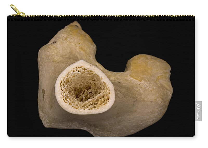
The upper (biting) surfaces of the tooth are at top, with the lower sections (bottom) embedded in the gums and jaw bone (not shown). There are trabeculae in spongy bone which gives its sponge like appearance. The central tubular region of the bone, called the diaphysis, flares outward near the end to form the metaphysis, which contains a largely cancellous, or spongy, interior. An outer 'fibrous layer' containing mainly fibroblasts, and an inner 'cambium layer' containing progenitor cells. Browse 53 bone marrow cross section stock photos and images available, or search for bone cross section or bone cells to find more great stock photos and pictures.
Bone Cross Section Lm Carry All Pouch For Sale By Science Stock Photography from render.fineartamerica.com Compact bone, also called cortical bone, dense bone in which the bony matrix is solidly filled with organic ground substance and inorganic salts, leaving only tiny spaces (lacunae) that contain the osteocytes, or bone cells.compact bone makes up 80 percent of the human skeleton; The diaphysis is the tubular shaft that runs between the proximal and. The surface features of bones vary considerably, depending on the function and location in the body. There are trabeculae in spongy bone which gives its sponge like appearance. Describes the bone markings, which are illustrated in (). The surface features of bones vary considerably, depending on the function and location in the body. Cross‐sectional area is derived from the integral of the bone mass profile across the narrow region. To cover the whole cortex the average area of acquisition was 4.3 × 4.0 mm.
After a fracture, woven bone forms initially and is gradually replaced by lamellar bone during a process known as bony substitution.
ads/bitcoin2.txt
The compact bone is made up of osteon. There are three general classes of bone markings: If you look at the cross section of a long bone under a microscope, the rings of bone immediately internal to the periosteum of the bone are called external circumferential lamellae. And why does the marrow stop where it does, and so sharply? The remainder is cancellous bone, which has a spongelike appearance with numerous large spaces and is found in the. Muscles and bones of the human body 12 photos of the muscles and bones of the human body anatomy bones of the human body quiz, major muscles and bones in the human body, muscles and bones in the human body, number of muscles and bones in the human body. Bone matrix and cells bone matrix osseous tissue is a connective tissue and like all connective tissues contains relatively few cells and large amounts of extracellular matrix. Describes the bone markings, which are illustrated in (). While it is not as hard as compact bone, spongy bone plays an important role of protecting the marrow where blood cells are produced. Bone structure right foot 12 photos of the bone structure right foot bone structure in. Would it be a good thing to show the epiphyseal plate? The surface features of bones vary considerably, depending on the function and location in the body. Compact bone is the outer layer and the spongy bone forms the.
This is known as the periosteum. Select from premium cross section of bone of the highest quality. To cover the whole cortex the average area of acquisition was 4.3 × 4.0 mm. Compact bone is very different from the other tissues you have seen. As the names suggest compact bone looks compact and the spongy bone looks like sponges.
File Bone Cross Section Svg Wikimedia Commons Bone Marrow Yellow Bone Marrow Cancellous Bone from i.pinimg.com Table 1 describes the bone markings, which are illustrated in (figure 4). Spongy bone and compact bone. The surface features of bones vary considerably, depending on the function and location in the body. Marrow in the shaft of long bones is typically yellow, with red marrow in the head through the cancellous bone. Learn vocabulary, terms, and more with flashcards, games, and other study tools. Compact bone, also called cortical bone, dense bone in which the bony matrix is solidly filled with organic ground substance and inorganic salts, leaving only tiny spaces (lacunae) that contain the osteocytes, or bone cells.compact bone makes up 80 percent of the human skeleton; Muscles and bones of the human body 12 photos of the muscles and bones of the human body anatomy bones of the human body quiz, major muscles and bones in the human body, muscles and bones in the human body, number of muscles and bones in the human body. Bone structure right foot 12 photos of the bone structure right foot bone structure in.
When modeled as a beam, a long bone's cross‐sectional geometry can provide measures of its compressive strength (e.g., cross‐sectional area csa and cortical area ca), as well as its resistance to bending and torsion about a particular axis (i.e., second moments of area i and polar moments of area j , respectively;
ads/bitcoin2.txt
Browse 53 bone marrow cross section stock photos and images available, or search for bone cross section or bone cells to find more great stock photos and pictures. The surface features of bones vary considerably, depending on the function and location in the body. Slides have to be made this way because the matrix of bone is too hard to be cut with a knife as the other tissues are. When modeled as a beam, a long bone's cross‐sectional geometry can provide measures of its compressive strength (e.g., cross‐sectional area csa and cortical area ca), as well as its resistance to bending and torsion about a particular axis (i.e., second moments of area i and polar moments of area j , respectively; This section will examine the gross anatomy of bone first and then move on to its histology. Compact bone, also called cortical bone, dense bone in which the bony matrix is solidly filled with organic ground substance and inorganic salts, leaving only tiny spaces (lacunae) that contain the osteocytes, or bone cells.compact bone makes up 80 percent of the human skeleton; There are three general classes of bone markings: Bone markings the surface features of bones vary considerably, depending on the function and location in the body. Compact bone, spongy bone, and bone marrow. Internal structure of a human long bone, with a magnified cross section of the interior. There are trabeculae in spongy bone which gives its sponge like appearance. Muscles and bones of the human body 12 photos of the muscles and bones of the human body anatomy bones of the human body quiz, major muscles and bones in the human body, muscles and bones in the human body, number of muscles and bones in the human body. An outer 'fibrous layer' containing mainly fibroblasts, and an inner 'cambium layer' containing progenitor cells.
And why does the marrow stop where it does, and so sharply? The central tubular region of the bone, called the diaphysis, flares outward near the end to form the metaphysis, which contains a largely cancellous, or spongy, interior. The upper (biting) surfaces of the tooth are at top, with the lower sections (bottom) embedded in the gums and jaw bone (not shown). Marrow in the shaft of long bones is typically yellow, with red marrow in the head through the cancellous bone. The cortical bone equivalent area of the cross‐section of the region of interest (femoral neck or shaft), with all soft tissue voids (trabecular and cellular spaces) eliminated (cm 2).
Skeleton Anatomy Vector Photo Free Trial Bigstock from static1.bigstockphoto.com Find the perfect cross section of bone stock photos and editorial news pictures from getty images. In a cross section of a bone we can see two types of bone tissue: Would it be a good thing to show the epiphyseal plate? Find the perfect cross section bone stock photo. Why is the marrow red? This is known as the periosteum. The cortical bone equivalent area of the cross‐section of the region of interest (femoral neck or shaft), with all soft tissue voids (trabecular and cellular spaces) eliminated (cm 2). Compact bone is the outer layer and the spongy bone forms the inner layer.
There are three general classes of bone markings:
ads/bitcoin2.txt
Describes the bone markings, which are illustrated in (). Now that you know what bones do, let's take a look at what they're made of and their anatomy. Each bone in your body is made up of three main types of bone material: If you look at the cross section of a long bone under a microscope, the rings of bone immediately internal to the periosteum of the bone are called external circumferential lamellae. Spongy bone and compact bone. Compact bone is very different from the other tissues you have seen. Find the perfect cross section of bone stock photos and editorial news pictures from getty images. When modeled as a beam, a long bone's cross‐sectional geometry can provide measures of its compressive strength (e.g., cross‐sectional area csa and cortical area ca), as well as its resistance to bending and torsion about a particular axis (i.e., second moments of area i and polar moments of area j , respectively; After a fracture, woven bone forms initially and is gradually replaced by lamellar bone during a process known as bony substitution. The structure of a long bone allows for the best visualization of all of the parts of a bone. There are trabeculae in spongy bone which gives its sponge like appearance. Browse 53 bone marrow cross section stock photos and images available, or search for bone cross section or bone cells to find more great stock photos and pictures. Marrow in the shaft of long bones is typically yellow, with red marrow in the head through the cancellous bone.
ads/bitcoin3.txt
ads/bitcoin4.txt
ads/bitcoin5.txt
0 Response to "Cross Section Of A Bone / Cross Section Of Bone Histoanatomy Youtube"


0 Response to "Cross Section Of A Bone / Cross Section Of Bone Histoanatomy Youtube"
Post a Comment